Floaters and Flashes - Retinal Detachment.
The most common causes of floaters and flashes are vitreous syneresis and posterior vitreous detachment (PVD) of the retina. Retinal detachment happens as a normal part of aging. The vitreous gel shrinks and separates from the retina. PVD normally happens over a period of time, and it's something that you won't feel. We invite you
to call to have your questions answered or to make an appointment at East Valley Ophthalmology in Mesa, Arizona: 480-981-6111.
What are Floaters?
Floaters are deposits of various size, shape, and consistency floating within
the normally transparent fluid (vitreous) inside the eye, appearing as spots,
threads, or fragments of cobwebs singly or together with several others in
one's field of vision — especially when looking
at a blank surface or an open monochromal space, such as blue sky.
Despite the name "floaters",
many of these specks have a tendency to sink toward the bottom of the eyeball,
in whichever way the eyeball is oriented; looking up or
lying back tends to concentrate them near the the center of
one's gaze, while the textureless and evenly lit sky forms an ideal background
against which to view them .
Sometimes fine dark lines in amorphous mass - like small branching twigs -
are seen. These floaters move around and are called 'muscae volitantes'
(Latin for 'flying flies'), because they seem to dart about like
flies as the eye is moved. Over time, you will become less aware of these floaters
as the brain learns to ignore these retinal images. Therefore, while some floaters
may remain in your vision, many of them will fade over time and become less
bothersome.
The Location of Floaters
The vitreous is a normally clear, gel-like substance that fills the center
of the eye. It makes up approximately 2/3 of the eye's volume, giving it form
and shape before birth.

Floaters, when present, are suspended in the vitreous. Thus, they generally
follow the rapid motions of the eye, while drifting slowly within the fluid.
When they are first noticed, the natural reaction is to attempt to look directly
at them. However, attempting to shift one's gaze toward them can be difficult
since floaters follow the motion of the eye, remaining to the side of the direction
of gaze. Floaters are, in fact, visible only because they do not remain perfectly
fixed within the eye. Although the blood vessels of the eye also obstruct light,
they are invisible under normal circumstances because they are fixed in location
relative to the retina, and the brain "tunes out" stabilized images
due to neural adaptation. This does not occur with floaters and they remain
visible.
The shapes are shadows projected onto the retina by tiny
structures of protein or other cell debris discarded over the years and trapped
in the vitreous humour. Floaters can even be seen when the eyes are closed
on especially bright days, when sufficient light penetrates the eyelids to
cast the shadows.
The Occurrence of Floaters
Floaters are common
and do not cause problems for most people. In fact, people usually
learn to ignore them, given some time. However, to those with severe cases,
floaters are definitely a distraction — especially if the spots seem
to constantly drift through the field of vision.
Floaters have been known to catch and refract light in ways that somewhat
blur vision temporarily until the floater moves to a different area. Many times
they trick the sufferer into thinking they see something out of the corner
of their eye that really is not there. For
people with severe floaters, it is nearly impossible to completely ignore
the large masses that constantly stay within almost direct view. Some sufferers
have noted a decrease in ability to concentrate while reading, watching television,
walking outdoors, and driving, especially when tired.
Although more common in elderly people, floaters can certainly become a
problem to younger people, especially if they are myopic. Floaters are also
more common after cataract operations or after trauma. In some cases, floaters
are congenital.
Tear Film Debris
Sometimes the appearance of floaters has to be attributed to dark specks
in the tear film of the eye. Technically, these are not floaters, but they
do look the same from the viewpoint of the patient. People with blepharitis
or a dysfunctional meibomian gland are especially prone to this cause, but
ocular allergies or even the wearing of contact lenses can cause the problem.
To differentiate between material in the vitreous humour of the eye and debris
in the tear film, one can look at the effect of blinking: debris in the tear
film will move quickly with a blink, while floaters are largely unresponsive
to it. Tear film debris is diagnosed by eliminating the possibility of
floaters and macular degeneration.
What are Flashes?
Flashes of light lasting a few seconds may appear in your vision when the
vitreous gel pulls or tugs on the retina. This may happen as a natural result
of aging or it may occur temporarily if you receive a blow to the head or
eye. Usually these flashes, which are often described as lightning streaks,
are noticed at night.
Light flashes appearing as wavy lines in both eyes and lasting from a few
minutes to half-an-hour, are usually a sign of an ocular migraine headache.
Migraine-related flashes are often noticed in a lighted environment. Flashes
of this nature are not a symptom of eye problems. The onset of new light flashes
of short duration at night, especially when accompanied by the appearance
of many new floaters or a blackening out of part of your field of vision,
may indicate a retinal tear or detachment.

Retinal detachment requires immediate medical
attention, as it can easily cause blindness. Both the appearance of flashes
and the sudden onset of numerous small floaters warrant an ophthalmological
investigation.
Causes of Floaters
There are other causes for the appearance of floaters, of which
the most common are described here. Basically, any way by which material enters
the vitreous humour is a cause for floaters.
Vitreous syneresis
The most common cause of floaters is vitreous syneresis, shrinkage of the
vitreous humour.
|
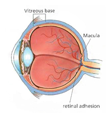
In the young eye, the vitreous gel is solid and the posterior vitreous
surface is well attached to the retinal surface.
|
|
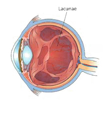
In the aging eye, small pockets of liquidfied vitreous (lacunae)
can develop within the gel.
|
|
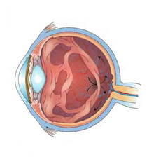
Lacunae may form pockets and lead to separation from
the retinal surface.
|
|
Contraction of the vitreous
can produce light flashes
or can cause physical tears.
|
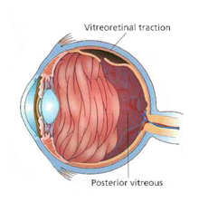
Posterior vitreous detachments and retinal detachments
Over time, as
we age, the vitreous loses support and its framework contracts. This may
lead to vitreous detachment, in which the vitreous body is released from
the retina. During this detachment, the shrinking vitreous can stimulate the
retina mechanically, causing the patient to see random flashes across the
visual field, sometimes referred to as "flashers."
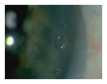
The ultimate release of the vitreous sometimes makes a large floater appear,
usually in the shape of a ring ("Weiss ring").
As a complication, part of the retina might be torn off by the departing
vitreous body, in a process known as retinal detachment. This will often
leak blood into the vitreous, which is seen by the patient as a sudden appearance
of numerous small dots, moving across the whole field of vision.

Regression of the hyaloid artery during pregnancy
The hyaloid artery, an artery running through the vitreous humour during the
fetal stage of development, regresses in the third trimester of pregnancy.
Its disintegration can sometimes leave cell matter.
Other common causes
Patients with retinal tears may experience floaters if red blood cells are
released from leaky blood vessels, and those with a posterior uveitis or vitritis,
as in toxoplasmosis, may experience multiple floaters and decreased vision
due to the accumulation of white blood cells in the vitreous humour.
Other causes for floaters include cystoid macular edema and asteroid hyalosis.
The latter is an anomaly of the vitreous humour, where by calcium clumps attach
themselves to the collagen network. The bodies that are formed in this way
move slightly with eye movement, but then return to their fixed position.
Diagnosis of Floaters
Usually the appearance of new floaters or light flashes does not indicate
any serious eye problem. However, the only way to ensure that the floaters
or flashes are not symptomatic of a more serious problem, is to have your retina
examined. If, following the exam, you develop large numbers of new floaters
that seem to get worse over time, we recommend that you have your eyes re-examined.
Standard vision tests like the Snellen visual acuity measurement,
which measures your vision as 20/20, etc., are unable to quantify floaters
and how the disability interferes with day-to-day functioning and overall quality
of life. Floaters are often readily observed with the use of an ophthalmoscope
or slit lamp. However, if the floater is a small piece of debris and near the
retina, your eye doctor may not be able to observe it even if it appears large
and obvious to you.
Increasing background illumination or using a pinhole to effectively decrease
pupil diameter may allow a person to obtain a better view of his or her own
floaters. The head may be tilted in such a way that one of the floaters drifts
towards the central axis of the eye. In the sharpened image the fibrous elements
are more conspicuous. (If the pinhole is kept moving slowly in small circles,
the same technique evokes an interesting entoptic effect known as the vascular
figure, which is a view of the blood vessels within one's own eye.)
Treatment of Floaters
When floaters appear in your line of vision, move your eye around — up and
down as well as from side to side. This movement creates a swirling in the
vitreous fluid and may cause the floater to move out of your field of vision.
Regular Check-Ups
To safeguard your vision, individuals over age 40 should
undergo a comprehensive eye exam annually. If you are under age 40 and have
risk factors such as high blood pressure, diabetes or a family history of glaucoma
or macular degeneration, a yearly exam is also recommended.Individuals under
age 40 who are in good health, with no known risk factors should have their
eyes examined every two years.
In some patients
floaters can cause persistent, distracting and disabling symptoms. In these
patients, visual tasks such as reading or driving become laborious, and productivity
and overall quality of life may suffer. It is advisable to wait at least one
year to allow enough time for the floaters to become less prominent naturally.
If they do not, then one may consider vitrectomy surgery or laser surgery.
We must caution at the very outset of this discussion that laser or surgical
treatment of floaters is not considered a standard management strategy
and is recommended by a minority of physicians, even then under exceptional
circumstances. We are not aware of any vitamins or drugs that can reduce floaters.
Pars Plana Vitrectomy (PPV)
Pars plana vitrectomy is a procedure usually reserved for complicated
posterior segment disease. It has a well-known risk profile, and justifiably
there is reluctance to offer this surgery to treat floaters. However, the post-operative
complication rate following PPV has been assessed in the setting of retinal
detachment surgery or in the presence of complicated vitreo-retinal disease.
It may be argued that pars plana vitrectomy for floaters, in eyes that have
an established posterior vitreous detachment, may be associated with
a lower incidence of both intraoperative and post-operative complications.
The three main postoperative complications that one must consider in vitrectomy
are: development or progression of nuclear sclerosis cataract, retinal detachment
and choroidal or vitreous hemorrhage (bleeding) in the eye.
The eye specialists of East Valley Ophthalmology perform advanced
technology diagnostic testing and treatment, as well as taking
the time necessary to provide each patient with information needed
to fully understand their condition and to achieve their best possible
visual outcome.
If you would like further information, please call our office at:
480-981-6111
East Valley Ophthalmology
Eye Doctors - Mesa, ArizonaIf you or a family member
or friend have not had a recent routine eye examination, have a specific eye condition that needs addressing, or are looking for
an eye specialist or professional eye consultant please take a moment to Request an Appointment.
|